‘It opens up a whole new universe’: Revolutionary microscopy technique sees individual atoms for first time
Cryo-electron microscopy breaks a key barrier that will allow the workings of proteins to be probed in unprecedented detail.

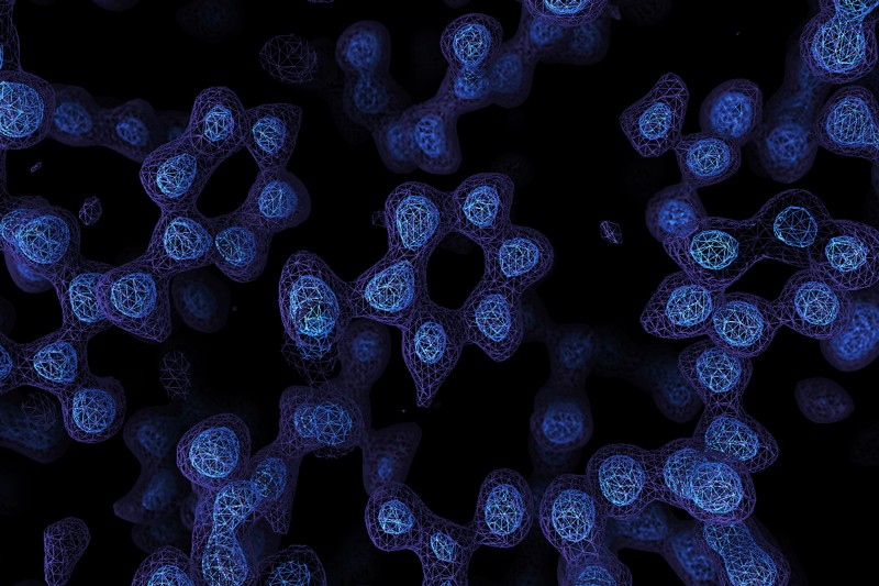
A Cryo-EM map of the protein apoferritin.Credit: Paul Emsley/MRC Laboratory of Molecular Biology
A game-changing technique for imaging molecules known as cryo-electron microscopy has produced its sharpest pictures yet — and, for the first time, discerned individual atoms in a protein.
By achieving atomic resolution using cryogenic-electron microscopy (cryo-EM), researchers will be able to understand, in unprecedented detail, the workings of proteins that cannot easily be examined by other imaging techniques, such as X-ray crystallography.
The breakthrough, reported by two laboratories late last month, cements cryo-EM’s position as the dominant tool for mapping the 3D shapes of proteins, say scientists. Ultimately, these structures will help researchers to understand how proteins work in health and disease, and lead to better drugs with fewer side effects.
“It’s really a milestone, that’s for sure. There’s really nothing to break anymore. This was the last resolution barrier,” says Holger Stark, a biochemist and electron microscopist at the Max Planck Institute for Biophysical Chemistry in Göttingen, Germany, who led one of the studies1. The other2 was led by Sjors Scheres and Radu Aricescu, structural biologists at the Medical Research Council Laboratory of Molecular Biology (MRC-LMB) in Cambridge, UK. Both were posted on the bioRxiv preprint server on 22 May.
“True ‘atomic resolution’ is a real milestone,” adds John Rubinstein, a structural biologist at the University of Toronto in Canada. Getting atomic-resolution structures of many proteins will still be a daunting task because of other challenges, such as a protein’s flexibility. "These preprints show where one can get to if those other limitations can be addressed,” he adds.
Breaking boundaries
Cryo-EM is a decades-old technique that determines the shape of flash-frozen samples by firing electrons at them and recording the resulting images. Advances in technology for detecting the ricocheting electrons and in image-analysis software catalysed a ‘resolution revolution’ that started around 2013. This led to protein structures that were sharper than ever before — and nearly as good as those obtained from X-ray crystallography, an older technique that infers structures from diffraction patterns made by protein crystals when they are bombarded with X-rays.
Subsequent hardware and software advances led to more improvements in the resolution of cryo-EM structures. But scientists have had to largely rely on X-ray crystallography for obtaining atomic-resolution structures. However, researchers can spend months to years getting a protein to crystallize, and many medically important proteins won’t form usable crystals; cryo-EM, by contrast, requires only that the protein be in a purified solution.
Atomic-resolution maps are precise enough to unambiguously discern the position of individual atoms in a protein, at a resolution of around 1.2 ångströms (1.2 × 10⁻¹⁰ m). These structures are especially useful for understanding how enzymes work and using those insights to identify drugs that can block their activity.
To push cryo-EM to atomic resolution, the two teams worked on an iron-storing protein called apoferritin. Because of its rock-like stability, the protein has become a test bed for cryo-EM: a structure of the protein with a resolution of 1.54 ångströms was the previous record.

The teams then used technological improvements to take sharper pictures of apoferritin. Stark’s team got a 1.25-ångström structure of the protein, with help from an instrument that ensures that the electrons travel at similar speeds before hitting a sample, enhancing the resolution of the resulting images. Scheres, Aricescu and their group used a different technology to fire electrons travelling at similar speeds; they also benefited from a technology that reduces the noise generated after some electrons career off the protein sample, as well as a more sensitive electron-detecting camera. Their 1.2-ångström structure was so complete, says Scheres, that they could pick out individual hydrogen atoms, both in the protein and in surrounding water molecules.
Stark reckons that melding the technologies could push resolutions to around 1 ångström — but not much further. “Below 1 Å is almost impossible to reach for cryo-EM,” he says. Obtaining such a structure with existing state-of-the-art technology would take “several hundred years of data recording and a non-realistic amount of compute power and data-storage capacities”, his team estimates.
See clearly
Scheres and Aricescu also tested their improvements on a simplified form of a protein called GABAA receptor. The protein sits in the membrane of neurons and is a target for general anaesthetics, anxiety medications and many other drugs. Last year, Aricescu’s team used cryo-EM to map the protein to 2.5 ångströms4. But with the new kit, the researchers attained a 1.7-ångström resolution, with even better resolution in some key parts of the protein. “It was like peeling off a blur over your eyes,” Aricescu says. “At this resolution, every half ångström opens up a whole universe.”
The structure revealed never-before-seen details in the protein — including the water molecules in the pocket where a chemical called histamine sits. “That is a gold mine for structure-based drug design,” says Aricescu, because it shows how a drug could displace the water molecules, potentially resulting in medications with fewer side effects.
An atomic-resolution map of GABAA, which isn’t as stable as apoferritin, would be a challenge, says Scheres. “I don’t think it’s impossible, but it would be very impractical,” because of the vast amount of data that would need to be collected. But other improvements, particularly in how protein samples are prepared, could pave the way for atomic-resolution structures of GABAA and other biomedically important proteins. Protein solutions are frozen on tiny grids made of gold, and alterations to these grids could hold proteins even stiller5.
“Everyone is very excited and amazed by the truly astounding level of performance demonstrated by the MRC-LMB and Max Planck groups,” says Radostin Danev, a cryo-EM specialist at the University of Tokyo. But he agrees that sample preparation is the field’s major challenge for more wobbly proteins. “Sub-1.5-Å, or even sub-2-Å, resolution performance will remain accessible for some time to only well-behaved samples,” he says.
The breakthroughs are likely to cement cryo-EM’s position as the go-to tool for most structural studies, says Scheres. Drug companies, which covet atomic-resolution structures, might be even more likely to turn to cryo-EM. But Stark thinks X-ray crystallography will retain some appeal. If a protein can be crystallized — and that’s a big if — it’s relatively efficient to generate structures of it bound to thousands of potential drugs in a short amount of time. But it can still take hours to days to generate enough data for extremely high-resolution cryo-EM structures.
“There are still pros and cons for each of the techniques,” says Stark. “People have published lots of papers and reviews that say these latest advances in cryo-EM will be the death signal for X-ray. I doubt that.”
Posted by Cláudio H. Dahne
Comentários
Postar um comentário